Description
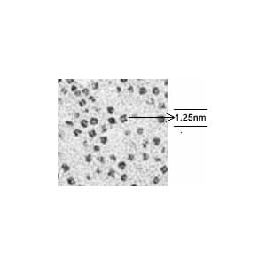
Determining resolution: Some ferritin molecules display a quad structure with a separation of 1.25nm. They are, therefore, useful as a resolution check. If a clear space can be resolved between two adjacent (not diagonal) electron dense particles within a molecule, then it can be assumed that the electron microscope is resolving at least 1.25nm. A photomicrograph negative magnification of at least 100,000x is recommended for this resolution check.
Individual ferritin particles are scattered throughout the specimen. Aggregates of particles are also present.
Ideal ferritin particles can be found around the edges of these aggregates. The ferritin particles will first become clearly visible using the EM binocular microscope with a screen magnification of around 20,000x

Reviews
There are no reviews yet.