
source: Ashley Yeager
Dead baby chicks from farms began arriving by the dozens at the vet labs of North Dakota Agricultural College in Fargo in 1930. Chicken farmers also brought in their sick chicks, many of which were gasping for air. Others were listless and seemingly depressed, with drooping wings and emaciated bodies. In 20 years of lab and field research, vets Arthur Schalk and Merle Hawn had never seen a chicken disease quite like this one. It ripped through poultry farms in North Dakota and Minnesota that year, killing tens of thousands of baby birds. Based on necropsies of the dead birds, the vets ruled out laryngotracheitis, commonly called the croup, as a cause of death (JAVMA, 78:413–22, 1931). In that disease, lesions appear in the windpipe, but in these chicks, tissue damage was found farther down, deep in the lungs. The illness was a new one, and Schalk and Hawn named it infectious bronchitis of baby chicks.
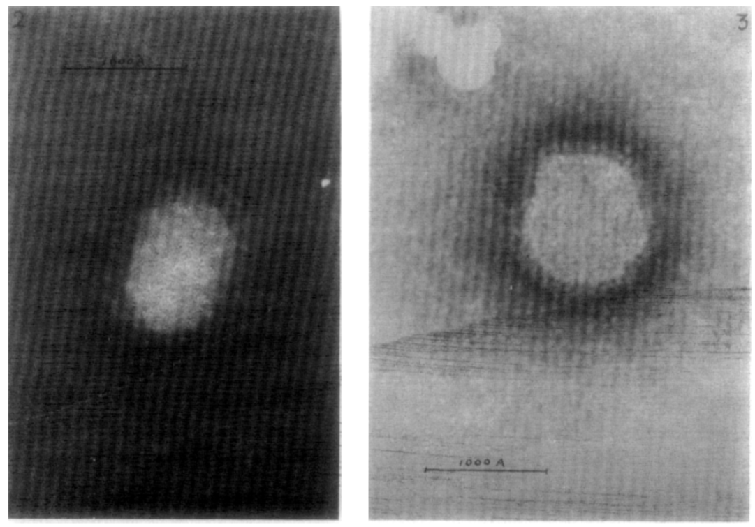
Alarmed at the impact on the poultry industry, other researchers began to investigate the disease, first identifying it as viral and naming the pathogen infectious bronchitis virus (IBV), and later revealing that its genetic code was RNA-based. It wasn’t until the 1960s that pathologists were able to get a look at the virus by staining the background of an electron microscope slide while leaving the actual specimen untouched, a technique called negative staining electron microscopy that was just emerging. Donald Berry of Glaxo Research, then located in a suburb of London, and his colleagues used the technique to capture fuzzy black-and-white images of an IBV particle and of an influenza A particle from a bird.
As Berry and his colleagues stared at the images, the researchers could see spikes jutting out of each viral particle. But the two types of viruses had different kinds of spikes. Influenza A’s were rod-shape and densely packed, while IBV’s were lollipop-shape, with bulbous growths at the ends, and they were much more spread out across the virus’s surface. IBV, Berry and his colleagues concluded in Virology in 1964, was not a form of the flu, as many had speculated. It was something else entirely.
Early negative staining electron microscopy images of viruses such as those in the 1964 paper offered the “next big breakthrough” in virology after identifying whether families of viruses were RNA- or DNA-based, says Jeffrey Kahn, a University of Texas Southwestern Medical Center pediatrician and infectious disease researcher who has written about the history of coronavirus research. Such early images gave clues about the true nature of viruses a decade before genetic sequencing became available.
About a year after Berry’s publication, June Almeida of the St. Thomas’s Hospital Medical School in London identified another virus in the same family as IBV—one that infected humans. Looking through an electron microscope at virus particles isolated from samples from a boy suffering flulike symptoms in Surrey, England, she too noticed the virus had lollipop-like protrusions, which she’d seen before on IBV and other viral particles she’d studied. Drawing on the resemblance between the fringe of viral spikes and the halo of gas, or corona, surrounding the sun, Almeida and others, including Berry, wrote to Nature in 1968 to characterize IBV and other recently discovered pathogens like it as a new viral family called coronaviruses.