Visualizing whole cells at many scales

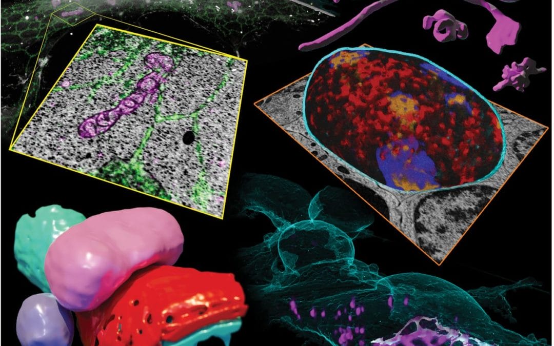
Cells need to compartmentalize thousands of distinct proteins, but the nanoscale spatial relationship of many proteins to overall intracellular ultrastructure remains poorly understood. Correlated light and electron microscopy approaches can help. Hoffman et al. combined cryogenic super-resolution fluorescence microscopy and focused ion beam–milling scanning electron microscopy to visualize protein-ultrastructure relationships in three dimensions across whole cells. The fusion of the two imaging modalities enabled identification and three-dimensional segmentation of morphologically complex structures within the crowded intracellular environment. The researchers observed unexpected relationships within a variety of cell types, including a web-like protein adhesion network between juxtaposed cerebellar granule neurons.
source Science Journal
