The Scientist Behind Some of the World's Best Coronavirus Images

From her laboratory in the far western reaches of Montana, Elizabeth Fischer is trying to help people see what they’re up against in COVID-19.
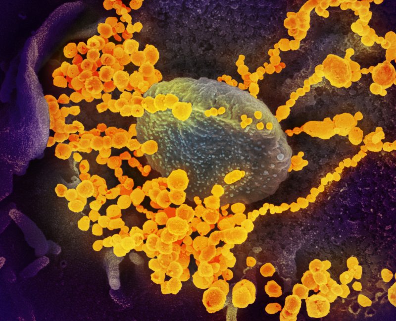
Over the past three decades, Fischer, 58, and her team at the Rocky Mountain Laboratories, part of the National Institutes of Health’s (NIH) National Institute of Allergy and Infectious Diseases, have captured and created some of the more dramatic images of the world’s most dangerous pathogens.
“I like to get images out there to try to convey that this is an entity, to try to demystify it, so this is something more tangible for people,” says Fischer, one of the country’s leading electron microscopists.
Now, as her renderings of the coronavirus flash across screens worldwide, she says: “You often hear people call it the invisible enemy. It’s trying to put that face out there.” Working in one of the nation’s 13 “Biosafety Level 4” labs—those equipped to safely handle the most dangerous pathogens—Fischer and her team visualize the world’s deadliest plagues from Ebola to HIV, salmonella to SARS-CoV-2, the coronavirus that causes COVID-19.


